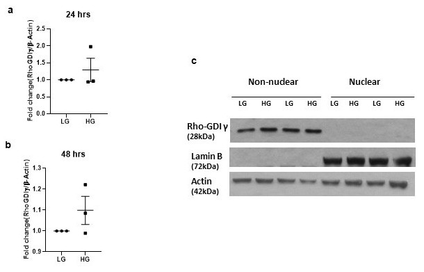
Fig. 4. Unlike RhoG, expression and subcellular distribution of GDIγ are not affected by hyperglycemic conditions. INS-1 832/13 cells were exposed to either basal glucose (LG, 2.5 mM) or high glucose (HG, 20 mM) for 24 (Panel a) or 48 (Panel b) hours. Relative abundance of RhoG in lysates was determined by Western blotting and band intensities were quantified by densitometry. Data are from three determinations in each condition. Panel c: INS-1 832/13 cells were exposed to either basal glucose (LG, 2.5 mM) or high glucose (HG, 20 mM) for 24 hours. Following incubation, nuclear and non-nuclear fractions were isolated, and relative abundance of Rho-GDIγ was determined in these fractions by Western blotting. Lamin B was used as marker protein for the nuclear fraction. Data from two individual experiments are shown here.